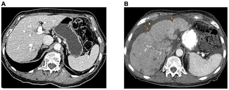
Figure 4:

Liver images of a single patient over time. Normal liver with subsequent progression to pseudocirrhosis
A: 8/14/09; smooth surface contour. B: 1/3/11 areas of capsular retraction (arrows) and generalized more fine nodularity of liver surface contour. New ascites*
