Figure 2.
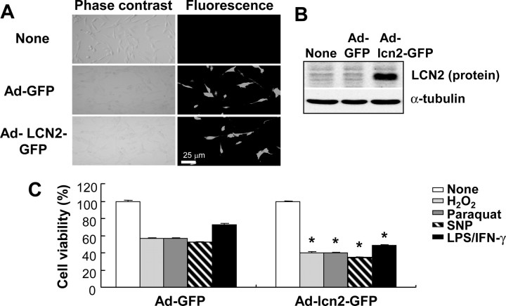
Augmented cell death in the astrocytes that were infected with adenoviral vector expressing lcn2. C6 glial cells were infected with adenoviral vectors expressing GFP (Ad-GFP) or lcn2 fused with GFP (Ad-lcn2-GFP) for 2 d, and then observed under fluorescence microscope (A). Magnification, ×200. Overexpression of lcn2-GFP fusion protein was confirmed by Western blot analysis using rabbit polyclonal anti-LCN2/NGAL antibody in the virus-infected cells (B). Virus-infected cells were treated with cytotoxic agents such as H2O2 (1 mm), paraquat (100 μm), SNP (0.5 mm), or a combination of LPS (100 ng/ml) and IFN-γ (50 U/ml) for 24 h, and then cell viability was assessed by MTT assay (C). The asterisks indicate statistically significant differences from the GFP-expressed cells (Ad-GFP) exposed to the same stimulus (p < 0.05; Ad-GFP vs Ad-lcn2-GFP). Values represent mean ± SD.

