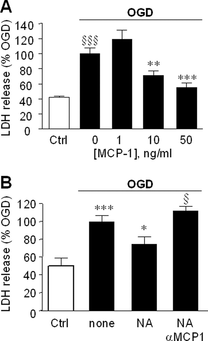
Figure 4.

MCP-1 reduces ischemic damage. A, Primary neurons were preincubated with the indicated concentrations of MCP-1 for 24 h, then exposed to OGD or kept under normoxic conditions [control (Ctrl); open bar], after which the media was replaced with fresh media (containing the same amounts of MCP-1). Neuronal viability was assessed 48 h later by measurement of LDH release. Data are mean ± SE of n = 12 replicates and shown relative to LDH release attributable to OGD alone. §§§p < 0.0005 versus control cells; **p < 0.005, ***p < 0.0001 versus OGD. B, Primary astrocytes growing on transwell membranes were transferred to wells containing primary neurons. After 24 h, the cocultures were treated for a further 24 h with fresh media (none), 10 μm NA (NA), or 10 μm NA and 10 μg/ml of MCP-1 neutralizing antibody (NA+αMCP1). After treatment, the astrocytes were removed, the media replaced, the neurons were exposed OGD, and neuronal viability assessed after 24 h. Control neurons (Ctrl; open bar) were kept normoxic. The data are the mean ± SE on n = 8 replicates and is LDH release relative to OGD treated only cells. ***p < 0.0005 versus control cells; *p < 0.05 versus none; §p < 0.05 versus NA. In control cells, 100% LDH reflected 20 ± 1.4% of total LDH released after 48 h (mean ± SE of n = 15 individual measurements).
