Figure 2.
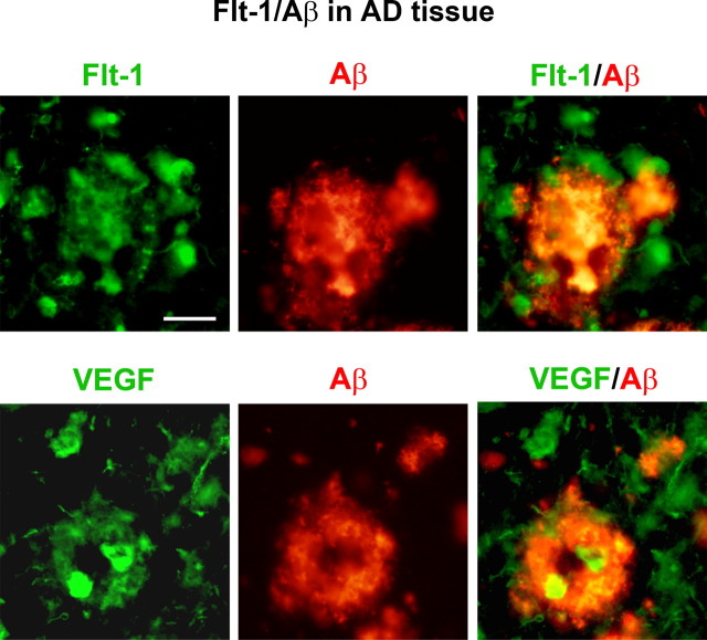
Association of Flt-1 and VEGF with β-amyloid peptide in AD tissue. The upper panels show representative images of Flt-1 and Aβ ir (left and middle panels) and merged staining of Flt-1/Aβ (right panel) in AD brain tissue. The lower panels show representative results for staining of VEGF with Aβ. Scale bar: 50 μm.

