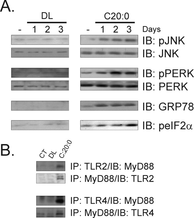
Figure 3.
A, Immunoblot (IB) analysis of the expressions of pJNK, JNK, pPERK, PERK, GRP78, and peIF2α in hypothalamic protein extracts obtained from rats treated intracerebroventricularly with diluent (DL) or arachidic acid (C20:0) for 1–3 d; some rats were intracerebroventricularly cannulated but received no treatment (−). B, Immunoprecipitation (IP) analysis of the associations of TLR2 and TLR4 with MyD88 in hypothalamic protein extracts obtained from nonintracerebroventricular cannulated (CT) rats, and rats treated intracerebroventricularly with DL or C20:0 for 3 d. The depicted blots are representative of five distinct experiments.

