
Figure 7.
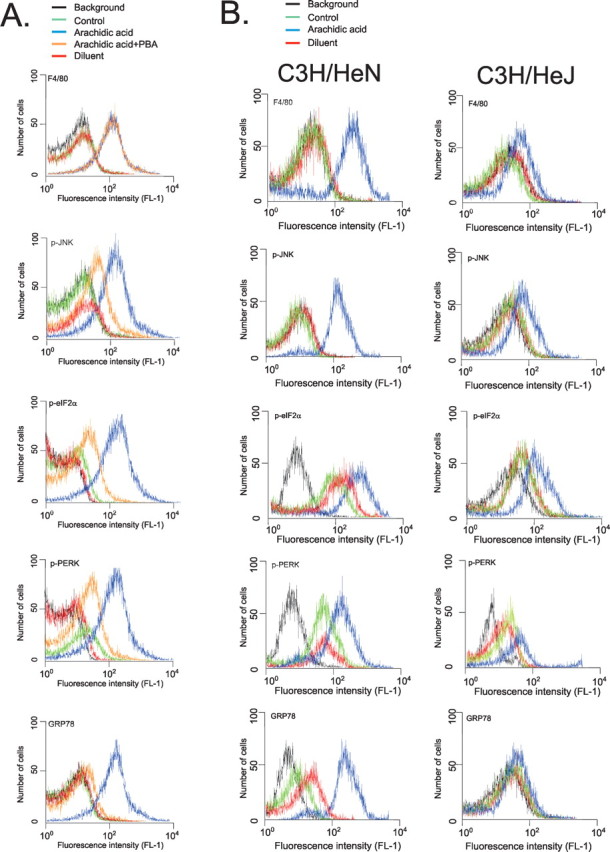
Flow cytometry analysis of expressions of F4/80, p-JNK, p-elf2α, p-PERK, and GRP78 in isolated macrophages. A, Macrophages from C3H/HeN mice were plated and incubated for 16 h in the presence of arachidic acid (blue), arachidic acid plus PBA (orange) or diluent (red). After harvesting, the cells were incubated with specific primary antibodies and then labeled with secondary conjugated antibody. Signal detection was performed by flow cytometry. B, Macrophages from C3H/HeN and C3H/HeJ mice were plated and incubated for 16 h in the presence of arachidic acid (blue) or diluent (red). After harvesting, the cells were incubated with specific primary antibodies and then labeled with secondary conjugated antibody. Signal detection was performed by flow cytometry. Graphs are representative of n = 5. Background counts (black); control, not added primary antibody (green).