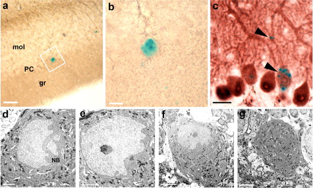
Figure 3.
Electron microscopic confirmation of single nuclei in LacZ-labeled Purkinje neurons in vav-iCre/LacZ mice. a, Example of a LacZ-positive Purkinje neuron, detected by X-gal staining of a 50-μm-thick Vibratome section, located in the Purkinje neuron layer (PC), between molecular (mol) and granule (gr) cell layer. b, Larger image of the same cell as in a. c, Light microscopic staining of calbindin confirming that all large X-gal-positive cell bodies in the PC are indeed Purkinje neurons. Typical dotted X-gal stain in the cell body and dendrite indicated by black arrowheads. d, e, Serial ultrathin sections through the same PKN as shown in a and b reveal only a single nucleus. The nucleus is large and light with a dark basophilic nucleolus, with deep folds and invaginations toward the side of the dendrites and Nissl bodies (NB) at the side of the folds, thus displaying the typical morphologic features of Purkinje neurons. f, g, Same cell at different z-levels and smaller magnification showing no second nucleus in this neuron. Scale bars: a–c, 20 μm; d, e, 2 μm; f, g, 5 μm.

