
Figure 7.
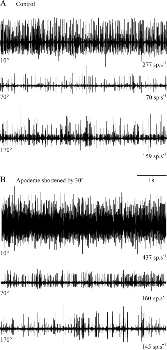
Extracellular recording of the activity of FCO sensory neurons at different angular positions of the femoro-tibial joint before (A) and after (B) apodeme shortening. All traces are from a single animal. The angular position of the tibia is indicated at the left of each trace and the mean spike frequency at the right. Shortening the apodeme caused an increase in activity at flexed joint angles (e.g., 10 and 70°) but not at extended joint angles (e.g., 170°).