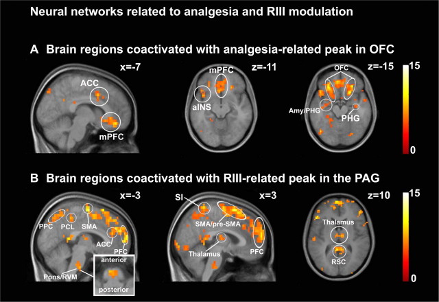
Figure 7.
Functional connectivity analysis. A, Brain regions coactivated with the analgesia-related peak in OFC. B, Brain regions coactivated with the RIII-modulation peak in the PAG. The inset is an enlargement of the RVM area. See Table 4, Regions coactivated with analgesia-related OFC and Regions coactivated with RIII-modulation peak in the PAG, respectively, for peak T values. PPC, Posterior parietal cortex; RSC, retrosplenial cortex.

