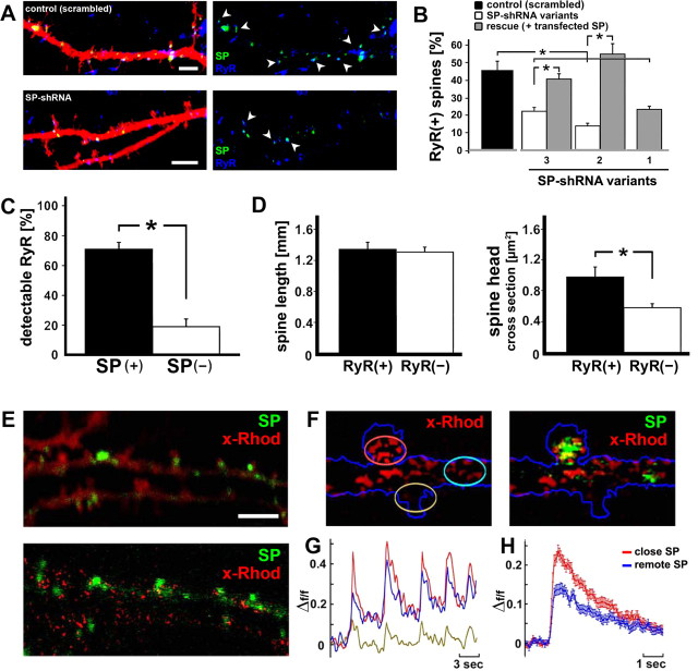
Figure 6.
SP is coupled to ryanodine receptors (RyR). A, Association between SP (green) and RyR (blue) in DsRed-transfected neurons. Both signals were frequently found adjacent to and touching each other within the same spines (see also supplemental Fig. 1, available at www.jneurosci.org as supplemental material). Arrowheads point at SP(+) spines. Scale bars, 2 μm. B, SP-shRNA-transfected neurons express fewer spine-associated RyR compared with cells transfected with a scrambled RNA plasmid (n = 3 cells per group; p < 0.001). The effects of shRNA variant 1, targeting the coding sequence of SP, cannot be rescued with cotransfected SP (cds only). C, D, RyR are more prevalent in SP(+) than in SP(−) spines (n = 6 cells, 101 spines; p < 0.01). D, No difference in spine length between RyR-positive [RyR(+)] and RyR-negative [RyR(−)] spines was seen (p > 0.895). The presence of RyR was associated with a larger spine head size (p < 0.02). E–H, Pulsed application of caffeine caused a transient rise of [Ca2+]i, nearer to SP puncta than more remote from them. Red indicates x-Rhod staining for imaging of [Ca2+]i, green GFP-SP. E, Raw image (top) and a subtracted image (bottom), indicating loci where x-Rhod shows enhanced fluorescence after exposure to caffeine. F, Regions of interest drawn near and remote from SP puncta. G, Illustration of responses to five consecutive pulse applications of caffeine; the individual traces correspond in color to those circled in F. H, Summary diagram of the time course of calcium changes near and remote from SP puncta in 48 dendrite/spine segments. The difference between the groups is highly significant (p < 0.001). These results demonstrate that SP is correlated with a larger calcium transient in response to caffeine.

