
Figure 8.
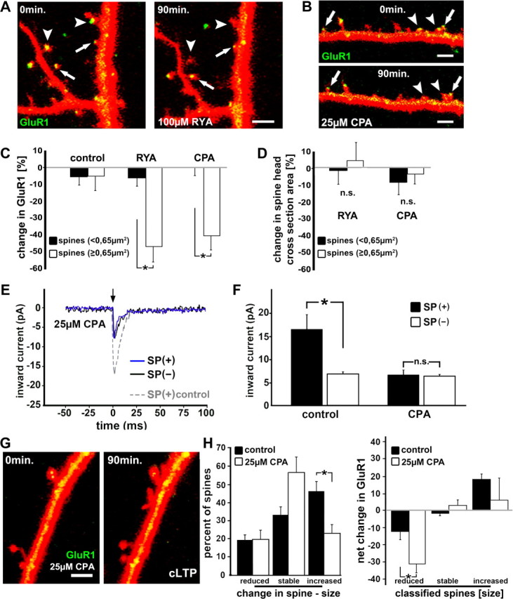
SP recruits RyR containing calcium stores to regulate GluR1 in dendritic spines. A, B, The density of GFP-GluR1 in DsRed-transfected cells is reduced after 90 min of exposure to 100 μm ryanodine or 25 μm CPA in spines with large heads (arrowheads) but not in spines with smaller heads (arrows), respectively. Scale bar, 1 μm. C, D, Summary diagram demonstrating the reduction of GFP-GluR1 fluorescent intensity in big spines in the presence of ryanodine or CPA (n = 5 cultures per group, five segments, one cell per culture; p < 0.01). Spine head size was not altered after 90 min in ryanodine or CPA (p > 0.2). E, Trace illustrations of inward current, generated at the flash (arrow; glutamate uncaging) demonstrate responses of SP(+) (blue trace) and SP(−) spines after 90 min of treatment with 25 μm CPA. Note the larger responses of an SP(+) spine under control conditions (gray dotted trace). F, The responses to the uncaging of glutamate in SP(+) spines is reduced down to the level seen in SP(−) spines after treatment with CPA (n = 4 cultures; four cells; 30 SP(+) spines; 26 SP(−) spines; four consecutive responses averaged per spine). G, H, The cLTP induction protocol was applied in CPA-treated cultures (25 μm) cotransfected with GFP-GluR1 and DsRed. Scale bar, 1 μm. Changes in spine size as well as changes in GluR1 fluorescence were compared before and 90 min after the induction of LTP. CPA-treated neurons (n = 4 cell, 180 spines) did not express a rise in GluR1 after the induction of LTP (p > 0.39), and significantly fewer spines were expanded after the induction of cLTP (H; p < 0.005).