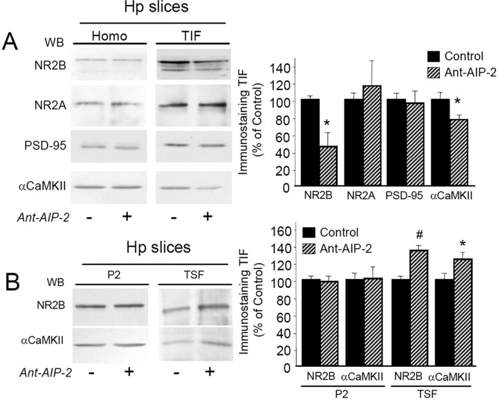
Figure 4.
Treatment with Ant-AIP-2 modulates NR2B localization in the postsynaptic fraction in acute hippocampal slices. A, Western blot analysis of the homogenate or TIF obtained from control and Ant-AIP-2 (10 μm, 2 h)-treated hippocampal slices. Same amount of proteins was loaded in each lane. Ant-AIP-2 treatment leads to a decreased NR2B localization in the TIF leaving the total amount of NR2B unaltered. NR2A and PSD-95 immunostaining in the TIF were not affected. Notably, αCaMKII levels in the TIF were also decreased. The graph displays the results of Western blot analysis expressed as control percentage (NR2B, *p < 0.05; αCaMKII, *p < 0.05, Ant-AIP-2 vs Control). B, Crude membrane fractions (P2) and TSF from control and Ant-AIP-2 (10 μm, 2 h)-treated hippocampal slices were analyzed by Western blot analysis with NR2B and αCaMKII antibodies. The graph displays the results of Western blot analysis expressed as control percentage (NR2B, #p < 0.01; αCaMKII, *p < 0.05, Ant-AIP-2 vs Control).

