
Figure 2.
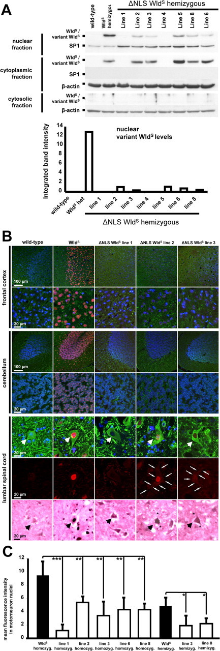
Reduced nuclear targeting of ΔNLS WldS in various neuronal subtypes in vivo. A, Western blots of nuclear, cytoplasmic, and cytosolic brain fractions demonstrating dramatically reduced nuclear targeting in individual mice from different ΔNLS WldS lines compared with spontaneous WldS mutant. The bottom graph demonstrates corresponding relative optic densities of nuclear WldS bands normalized to SP1 for individual mice. These Western blots are representative of three individual experiments. B, Rows 1–6, Confocal images showing (variant) WldS protein immunolabeling (red) with β-III tubulin (green) and Hoechst 33258 (blue) counterstaining on frozen sections from homozygous mice. Transgenics shown represent lines without detectable variant WldS protein expression in Western blotting (line 1) and with strong (line 2) and medium (line 3) variant WldS protein expression levels. Row 7, Light microscopic pictures from lumbar motorneurons (arrows) showing WldS immunolabeling using 3,3′-diaminobenzidine. C, Graph showing normalized fluorescence intensities for nuclear variant WldS protein signals in lumbar motoneurons. For each genotype group, three mice were assessed (10 motorneurons each). *p < 0.05; **p < 0.005; ***p < 0.001; one-way ANOVA.