
Figure 9.
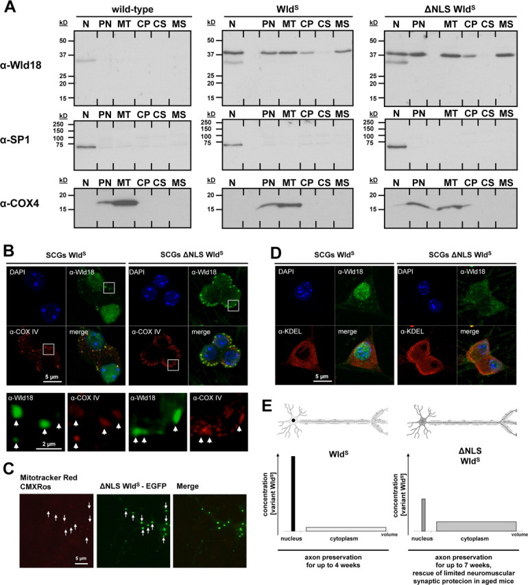
WldS and variant ΔNLS WldS proteins associate with mitochondria and microsomes. A, Western blot of WldS variants in subcellular fractions from mouse brain: N, nuclear; PN, postnuclear; MT, mitochondrial; CP, cytoplasmic; CS, cytosolic; MS, microsomal. Nuclear SP-1 and mitochondrial marker COX-IV were used to assess the fractionation. B, Confocal images of SCG neurons labeled with Wld18 (green; Alexa488-tyramide signal amplification) and COXIV (red) antibodies. White squares represent higher magnification insets shown in lower panel. Arrows indicate partial colocalization of Wld18 and COXIV. C, Confocal live cell imaging of hippocampal neuron expressing variant ΔNLS WldS fused to EGFP (green). The preparation was labeled with Mitotracker Red CMXRos (red). Arrows indicate partial colocalization of ΔNLS WldS-EGFP and mitochondria. D, Confocal images of SCG neurons labeled with Wld18 (green; Alexa488-tyramide signal amplification) and KDEL (red; as ER marker) antibodies. Note many extranuclear variant WldS foci overlap with the KDEL endoplasmic reticulum marker signals. E, Schematic illustration summarizing non-nuclear-mediated delay of Wallerian degeneration: altered ratio of nuclear versus cytoplasmic WldS content results in more robust neuroprotection in ΔNLS WldS transgenics. The graphs demonstrate distribution of WldS (left) with high concentration in the smaller nuclear compartment and low concentration in the much larger cytoplasmic compartment. In contrast, ΔNLS WldS (right) is altered in favor of the large cytoplasmic compartment resulting in easier detectability and in more robust axon and synapse protection.