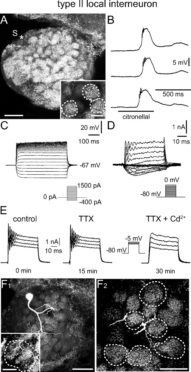
Figure 4.

Type II LN: electrophysiological and morphological characteristics. A, Morphology of the recorded neuron. The position of the soma (S) that was lost during processing is marked (*). The neuron innervated all glomeruli and the distribution of neurites was similar in all glomeruli. This is demonstrated with the inset, in which three glomeruli are outlined that are fully contained in the image stack (70 μm). B, C, Odor stimulation (B) and injection of depolarizing current (C) did not induce overshooting action potentials. B, Odor stimulation induced a graded depolarization and not Na+-driven spikelets, but no overshooting Na+-driven action potentials. C, The neuron showed rectification during injection of large depolarizing currents, but never generated overshooting, Na+-driven action potentials. Current was injected for 500 ms from −400 to 1500 pA in 100 pA steps. D, Whole-cell recording of (mainly) voltage-activated currents in normal saline. Depolarizing voltage steps from a holding potential of −80 mV did not elicit transient, TTX-sensitive Na+ currents. Voltage steps to membrane potentials to between −55 and −25 mV revealed an inactivating Ca2+ current. More depolarizing voltage steps to membrane potentials between −20 and 0 mV evoked outward K+ currents with 4-AP- and TEA-sensitive components. E, The oscillations that occurred at the beginning of these voltage steps were insensitive to high concentrations of TTX (10−4 m), but could be blocked by Cd2+ (5 × 10⁻⁴ m). F1, Image of another type II LN demonstrating the homogeneous innervation of each glomerulus (90 μm image stack). The inset shows a different z-stack demonstrating the innervation of the macroglomerulus (90 μm image stack). F2, Higher magnification substack from the neuron shown in E1 further demonstrates the homogenous glomerular innervation. The outlined glomeruli are fully contained in the image stack (70 μm). For abbreviations, see the legend of Figure 2. Scale bars: A, 100 μm; A inset, 20 μm; F1, 100 μm; F1 inset, F2, 50 μm.
