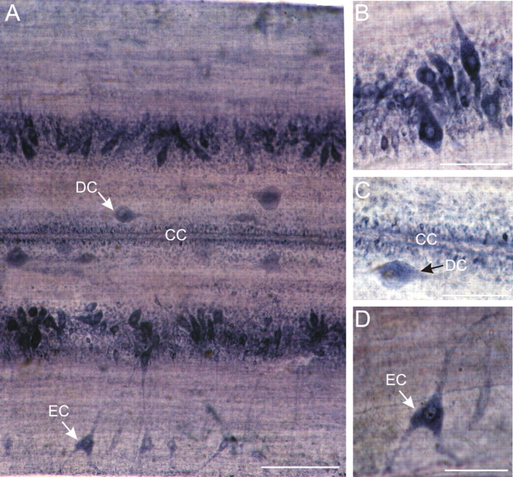
Figure 1.

NADPH-d labeling in the spinal cord of the lamprey. A, Whole-mount segment of spinal cord viewed dorsally. NADPH-d staining can be observed in different types of cell. Edge cells (EC) are located in the lateral columns and have long dendrites projecting toward edge of cord. Dorsal cells (DC) are large, rounded, and irregularly distributed along the medial columns. Ependymal cells surrounding the central canal (CC) are marked. Scale bar, 200 μm. B, Detail of a part of the gray column, which includes motoneurons and interneurons. C, Detail showing a dorsal cell (DC) medial to the central canal (CC). Note that the ependymal cells surrounding the central canal show NADPH-d reactivity. D, Enlargement of edge cell with its lateral dendrites. Scale bars: B–D, 100 μm.
