
Figure 8.
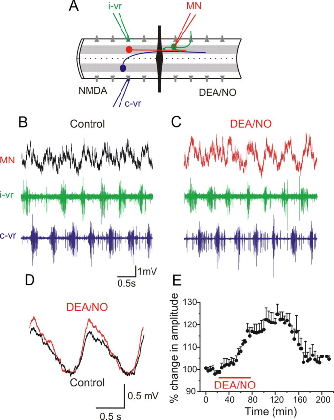
NO enhances the on-cycle excitation. A, Experimental arrangement with the recording chamber divided into two pools with an agar barrier. In the rostral part of the spinal cord locomotor activity was induced by NMDA while the caudal part was perfused with normal solution containing strychnine to block inhibition and intracellular recording was made from an MN. The red neuron represents ipsilateral excitatory interneurons and the blue neuron represents a commissural inhibitory interneuron. B, In control, the MN received excitation in phase with the ipsilateral ventral root (i-vr) burst. C, Application of DEA/NO (100 μm) in the caudal pool increased the amplitude of the excitatory synaptic input received by the MN. D, Averaged excitatory synaptic input received by the MN showing an increase in amplitude induced by DEA/NO. E, Plot of averaged data from a different experiment showing the time course of the change in the amplitude of the excitation before, during and after washout of DEA/NO (p < 0.0001, n = 8).