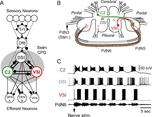
Figure 1.
The Tritonia swim network and swim motor program. A, A schematic diagram of the neural circuit underlying the swim motor pattern. All neurons have a contralateral counterpart that is not represented. The shaded circle indicates the neurons that comprise the swim CPG. The filled triangles represent excitatory synapses, and the filled circles represent inhibitory synapses. Combinations of triangle and circle are multicomponent synapses. The dotted lines indicate that the connection is either polysynaptic or not determined. C2, Cerebral cell 2; DFN-A,B, dorsal flexion neurons; DRI, dorsal ramp interneuron; DSI, dorsal swim interneuron; Tr1, trigger neuron 1; VFN, ventral flexion neuron; VSI, ventral swim interneuron [based on Getting et al. (1980), Getting (1981), Hume and Getting (1982), Frost and Katz (1996), and Frost et al. (2001)]. B, Schematic drawing of a dorsal view of the Tritonia brain, showing the location of the swim CPG neurons and their axonal projections. DSI and C2 are located on the dorsal surface of the cerebral ganglion. VSI is located on the ventral side of the pleural ganglion. C2 and VSI project their axons through PdN6, whereas DSI projects through PdN5. The dotted lines indicate PdN5 was removed on isolation of the brain. C, An example of the swim motor pattern. Simultaneous intracellular recordings from three CPG neurons, C2, DSI, and VSI, and extracellular recording from PdN6 are shown. The bursting pattern was elicited by stimulation of the left pedal nerve 3 by applying voltages pulses (8 V, 1 ms) at 5 Hz for 3 s (starting at arrow).

