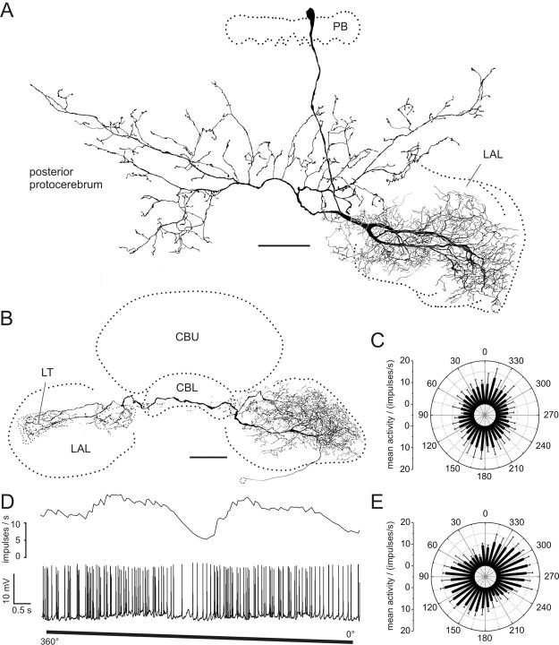
Figure 9.
POL neurons with input arborizations in the LAL. A, Frontal reconstruction of a LAL-pPC neuron. The cell has smooth endings in the left LAL. Bilateral projections to the posterior protocerebrum have varicose terminals. B, Frontal reconstruction of a LAL-LT neuron. The neuron connects the LAL of the left hemisphere with the LT in the right hemisphere. Side branches are also present in the right median olive. The location of the soma could only be inferred by the course of the faintly stained primary neurite. C, Circular plot of mean spiking frequency from the LAL-pPC neuron shown in A (means + SD, n = 6, bin size 10°, p = 4.2 × 10⁻⁸). D, Neuronal activity of the LAL-LT neuron presented in B during 360° rotation of the polarizer. E, Mean spiking activity of the LAL-LT neuron presented in B plotted against E-vector orientation during four rotations of the polarizer (error bars = SD, bin size 10°, p < 10−12). Scale bars, 100 μm.

