Figure 7.
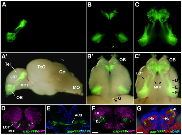
Preserved axon projection pattern of lhx2atg+ mitral cells in adult fish. A–C, A'–C', A whole-mount brain from lhx2a:gap-YFP adult fish is shown with lateral (A, A'), dorsal (B, B') and ventral (C, C') views. A–C, Fluorescence images. A'–C', Fluorescence superimposed on bright-field images. D–G, Coronal sections through the forebrain are immunostained with antibodies against gap-YFP (green), NPY, (magenta in D and F), and SV2 (red in G). Nuclear staining with DAPI (blue) is shown in E and G. Approximate positions of individual sections are indicated in B' and C'. Arrowheads in B' and G denote the gap-YFP+ axon terminals in the right habenula. Tel, Telencephalon; TeO, tectum opticum; Ce, cerebellum; MO, medulla oblongata; ACd, dorsal part of anterior commissure; lHb, left habenula; rHb, right habenula. Scale bar: in F, 200 μm for D–F; in G, 50 μm.

