
Figure 9.
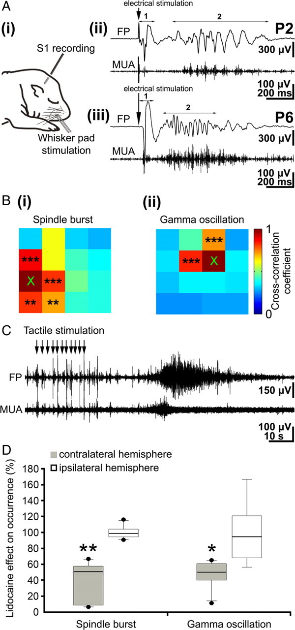
Stimulation of the periphery evokes neocortical oscillatory activity patterns. A, i, Scheme of experimental paradigm allowing electrical stimulation of the whisker pad and simultaneous recording of the field potential and MUA in the contralateral barrel cortex. ii, Field potential (top) and MUA (below) recordings of the contralateral S1 response to a single electrical stimulus of the whisker pad in a P2 rat. The robust direct response (1) is followed by a spindle burst (2) correlated with prominent MUA. iii, Field potential (top) and MUA (below) recordings of the contralateral S1 cortical response to electrical stimulation of the whisker pad in a P6 rat. The direct response (1) is followed by a gamma oscillation (2) correlated with MUA. B, Color-coded plot of maximal cross-correlation coefficients calculated for spindle bursts (i) and gamma oscillations (ii) elicited by electrical stimulation of the whisker pad. Asterisks indicate significant cross-correlation coefficients compared with the reference channel marked by ×. C, Field potential (top) and MUA (below) recordings of the contralateral S1 cortical response to repetitive tactile stimulation of the whiskers (11 times at ∼1 Hz) in a P6 rat. Repetitive stimulation of the sensory input elicits a robust long oscillation correlated with MUA. D, Effects of transient peripheral deafferentation by injection of lidocaine into the whisker pad on spontaneous spindle bursts and gamma oscillations. Box plots display the relative occurrence of spindle bursts (left) and gamma oscillations (right) recorded in six pups in the contralateral (black bars) and ipsilateral (white bars) barrel cortex after lidocaine injection.