
Figure 2.
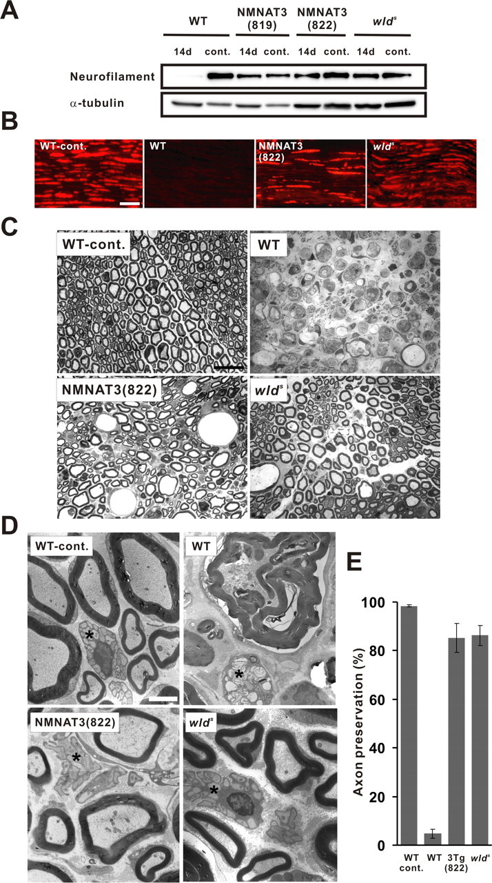
NMNAT3-Tg mice show delayed Wallerian degeneration comparable with wlds mice. Axonal degeneration was examined in a sciatic nerve injury model. Neurofilament immunoreactivity in the distal segment of the transected sciatic nerve was examined in NMNAT3-Tg (lines 819 and 822), wlds mice, as well as in wild-type control at 14 d after nerve lesion and compared with uninjured control by immunoblot analysis (A) and on longitudinal sections by immunohistochemistry 14 d after injury (B). α-Tubulin served as a loading control in A. Note that neurofilament expression remains close to wild-type level even at 14 d after injury. Scale bar, 50 μm. C, Injured nerve morphology in indicated mice was analyzed on toluidine blue-stained semithin sections (1 μm thick) 7 d after transection at 2 mm distal to the injury site. Injured nerves of NMNAT3-Tg and wlds mice exhibited apparently intact myelin structures almost comparable with uninjured control, whereas injured nerves in wild-type mice lacked intact myelin sheath and showed severe degeneration. Scale bar, 20 μm. D, Electron-microscopic images of the transverse sections of injured sciatic nerves at 2 mm distal to the transection site at 7 d after injury. Organized axonal structures and intact myelin sheath were observed in myelinated and unmyelinated (asterisk) axons of injured nerves in NMNAT3-Tg and wlds mice, whereas severe degeneration of both myelinated and unmyelinated (asterisk) axons and destruction of myelin structures was observed in injured nerves of wild-type mice. Scale bar, 2 μm. E, Morphologically preserved axon numbers in transverse sections of the injured sciatic nerve (7 d after transection) were counted for NMNAT3-Tg (line 822), wlds, and wild type mice (n = 7 for each genotype) and shown as percentage to the total number of axons. Error bars indicate SD.