Figure 6.

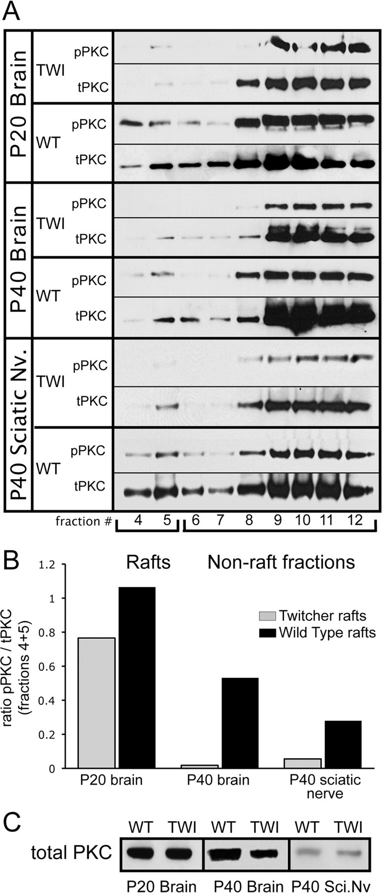
Psychosine accumulation parallels a decrease in protein kinase C in TWI lipid rafts. Lipid raft fractions from WT and TWI nervous tissue were analyzed using antibodies for total protein kinase C (tPKC) and phosphorylated PKC (pPKC). A, Western blots show the distribution of PKC and its active phosphorylated form in the brain at P20 and P40 and in the sciatic nerve at P40. These results show a decrease of total PKC in TWI rafts while also illustrating a much more significant loss of pPKC in these fractions. This indicates a significant reduction in the raft-based localization of PKC that occurs as psychosine concentrations go up in these membrane realms. B, Levels of activated (phosphorylated) PKC in raft fraction (4–5) are quantified with respect to the amount of total PKC in the same fractions. These results are expressed as a ratio of pPKC/tPKC. This analysis confirms a measurable reduction in pPKC in TWI tissues. C, Levels of PKC in total homogenates are presented from P20 and P40 brains and from P40 sciatic nerves.
