Figure 7.
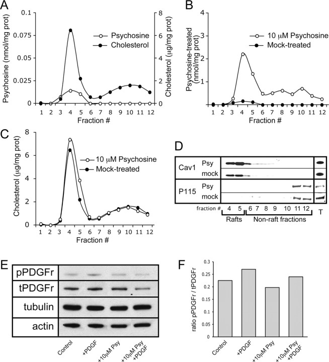
Treatment of cells in vitro with psychosine results in lipid raft alterations that mimic those observed in vivo. HeLa cells were cultured and treated with either 10 μm psychosine or vehicle for 24 h before being subjected to lipid raft preparation. A, Rafts from untreated HeLa cells were assayed for cholesterol levels, which are expressed in micrograms per milligram of protein, and endogenous psychosine levels which are expressed in nanomoles per milligram of protein. These analyses show an enrichment of both lipid species in raft fractions. B, Mass spectrometric detection in fractions from treated HeLa cells shows that exogenous addition of psychosine results in its targeted accumulation in rafts (fractions 4–5), which is expressed in nanomoles per milligram of protein. C, Analysis of cholesterol concentration in fractions from treated HeLa cells is expressed in micrograms per milligram of protein and shows an increase in cholesterol in the LR fractions (4–5) after psychosine exposure. D, The level and distribution of the raft marker protein caveolin-1 was assayed and shows an increase in abundance in raft fractions during treatment with psychosine. The nonraft marker P115 was also measured in addition to the measurement of the levels of both proteins in total cell lysates. E, Levels of phosphorylated and total PDGFr (pPDGFr and tPDGFr, respectively) along with levels of tubulin and actin were analyzed by Western blot. Analyses were done using HeLa cells in each of the following conditions: untreated control conditions (control), in the presence of PDGF (+PDGF), in the presence of 10 μm psychosine (+10 μm Psy), and in the presence of both PDGF and 10 μm psychosine (+10 μm Psy +PDGF). F, Quantification of the level of phosphorylation of PDGFr in E is presented. These results indicate that PDGFr can be activated even in the presence of psychosine. Measurements are presented as a ratio of pPDGFr/tPDGFr.

