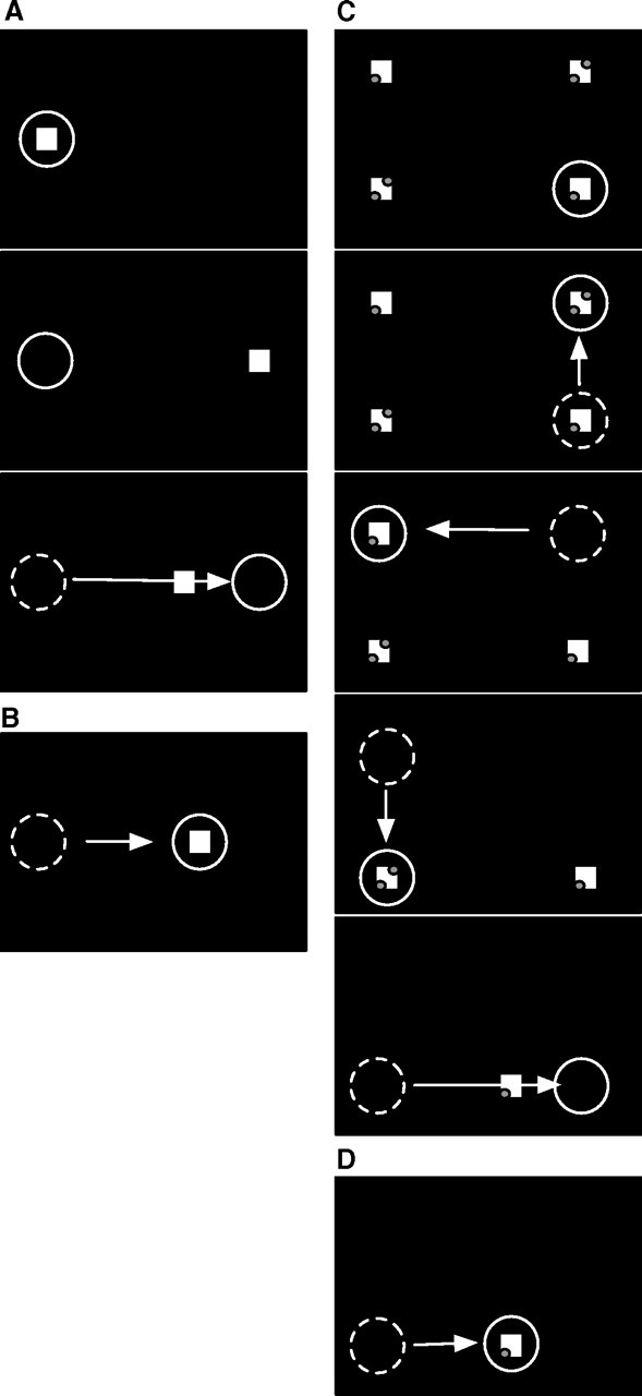
Figure 1.

A, Experimental procedure for reactive saccade adaptation. At the beginning of the trial (top panel), a fixation point (square) is presented near the left screen border. The subject's gaze (circle) is directed to the fixation point. After 1000 ms (middle panel), the fixation point disappears and a saccade target appears 30° to the right of the fixation point. The subject initiates the saccade to the target. When the saccade onset is detected (bottom panel), the saccade target is displaced, inducing a visual error after the saccade. B, After several such adaptation trials, the saccade amplitude becomes shorter. The saccade ends on the displaced target and the visual error after the saccade is reduced. C, Experimental procedure for scanning saccade adaptation. At trial onset (top panel), four saccade targets (squares) are presented. The subject fixates the bottom right target (circle). At a voluntary pace, the subject scans the targets in a counterclockwise manner. As the subject executes each saccade, the previously inspected target is extinguished. Adaptation takes place during the saccade from the bottom left to the bottom right target (bottom panel). When the onset of the saccade is detected, the bottom right target is displaced to the left, inducing a visual error after the saccade. D, After several adaptation trials, the saccade amplitude is adapted to the displaced target location.
