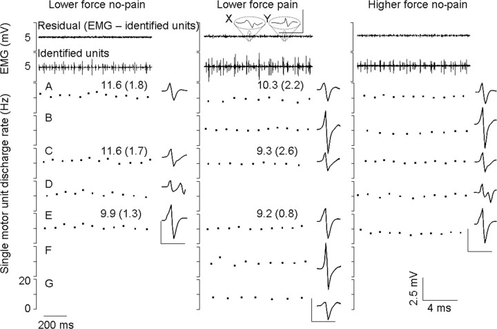
Figure 5.
Recordings of motor unit activity from a fine-wire electrode inserted into the FPL of one subject. Single motor units are shown with their respective discharge rates [mean (SD)] and spike-trigger averaged electrical profile. Once these units were identified and removed from the EMG trace, there was very little residual left (top trace). An exception of two small spikes in the residual of the painful contraction is highlighted (X and Y). Neither spike reflects a unit that regularly discharges, and therefore they do not meet the criteria as discussed in Materials and Methods. The small residual trace clearly indicates that all reliable units were identified from this file. Three motor units were recruited during both the no-pain and pain lower-force trials (A, C, and E). These units all decreased in discharge rate during pain. Three new units (B, F, and G) that were not seen in the lower-force no-pain conditions were recruited during pain. Of these units, only B is expected given orderly recruitment of the motoneuron pool as determined from those additional units recruited without pain at a slightly higher force level (B and D). Unit D is clearly not recruited during pain. The derecruitment of unit D, which coincides with the recruitment of B, F, and G during pain, demonstrates a change in recruitment during pain.

