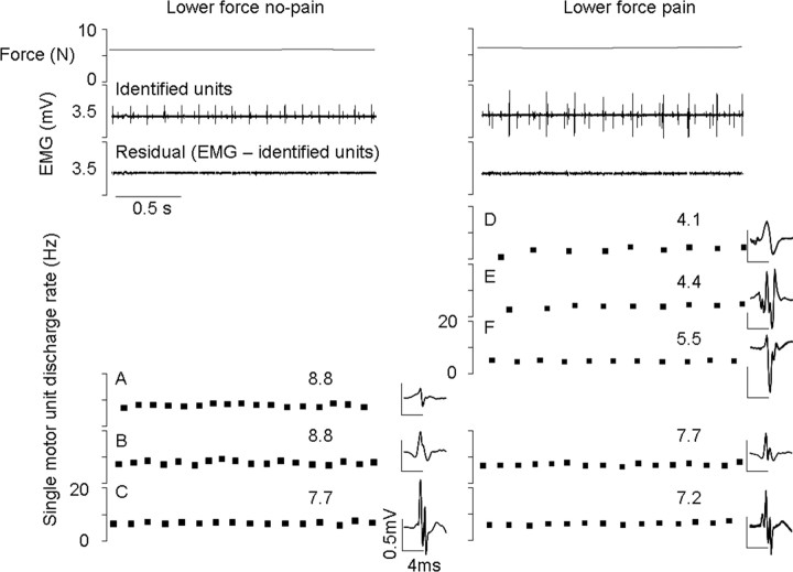
Figure 6.
Recordings of motor unit activity from a fine-wire electrode inserted into the quadriceps of one subject. Single motor units are shown with their respective discharge rates and spike-trigger averaged electrical profile. The small residual EMG trace clearly indicates that all reliable units were identified from this file. Two motor units were recruited during both the no-pain and pain lower-force trials (B and C); their motor unit firing rate decreased during pain. Three new units (D–F) that were not seen in the low-force no-pain conditions were recruited during pain. Unit A is not recruited during pain. The derecruitment of unit A, which coincides with the recruitment of D–F during pain, demonstrates a change in recruitment during pain.

