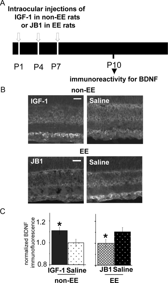
Figure 3.

IGF-1 expression controls BDNF protein levels in RGC layer at P10. A, Experimental protocol. B, Retinal section micrographs of P10 non-EE rats intraocularly injected with IGF-1 or saline (top) and of P10 EE rats intraocularly injected with JB1 or saline (bottom). BDNF-immunolabeled cells are detectable at the level of the RGC layer. Scale bars, 50 μm. C, Left, Quantitative analysis of mean BDNF immunofluorescence intensity in the RGC layer normalized to mean background level in the ONL layer of non-EE rats intraocularly injected with IGF-1 or saline and immunostained for BDNF at P10. BDNF immunoreactivity is significantly higher (asterisk) in the retinas of non-EE IGF-1-treated rats (1.103 ± 0.033; n = 7) with respect to those of non-EE saline-treated rats (1 ± 0.033; n = 7) (Mann–Whitney rank sum test; p < 0.001). Right, Mean BDNF immunofluorescence intensity in the RGC layer normalized to mean background level in the ONL layer of EE rats intraocularly injected with JB1 or vehicle and immunostained for BDNF at P10. BDNF immunoreactivity is significantly lower (asterisk) in the retinas of EE JB1-treated rats (1.001 ± 0.033; n = 7) with respect to those of EE saline-treated rats (1.104 ± 0.039; n = 5) (Mann–Whitney rank sum test; p < 0.001). Error bars represent SEM.
