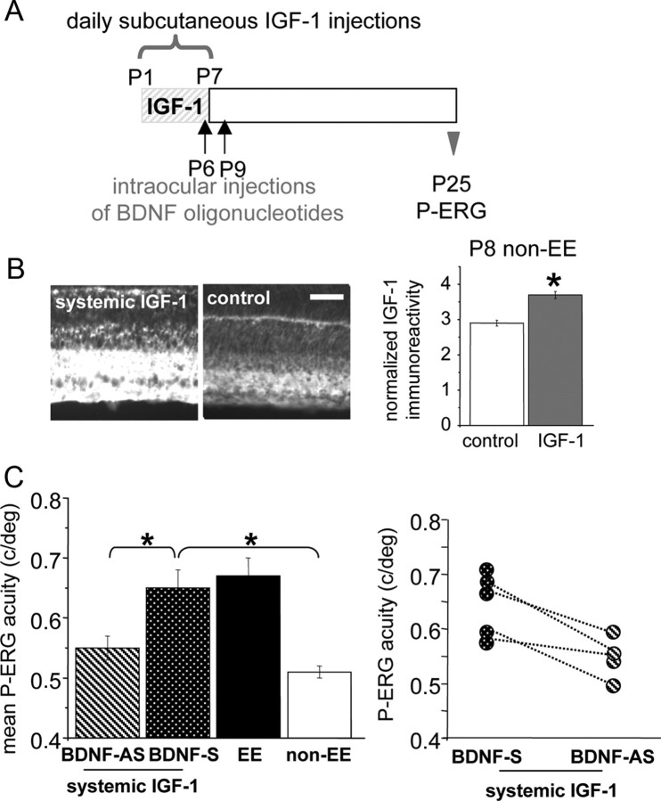
Figure 4.
Block of BDNF in the retina prevents the increase of retinal acuity present at P25 in IGF-1-treated rats. A, Schematic protocol of the experiment. B, Left, Representative images taken from P8 retinas processed for IGF-1 immunoreactivity. Right, Normalized IGF-1 expression in the RGC layer. IGF-1 levels in the RGC layer of IGF-1 systemically infused non-EE rats (3.72 ± 0.24; n = 4) are significantly higher (asterisks) than in control non-EE rats (2.92 ± 0.16; n = 4) (t test, p = 0.002). Scale bar, 50 μm. Error bars represent SEM. C, Left, Mean retinal acuity at P25 for IGF-1 systemically injected pups that had been intraocularly injected with BDNF-AS oligonucleotides (n = 4) in one eye and with BDNF-S oligonucleotides (n = 5) in the other. P-ERG acuity for the eye treated with antisense BDNF oligonucleotides is significantly lower [0.55 ± 0.02 cycles/degree (c/deg)] than in sense-treated eyes (0.65 ± 0.03 c/deg) or untreated eyes of EE animals (0.67 ± 0.03 c/deg; n = 5) and does not differ from the P-ERG acuity of P25 non-EE animals (0.51 ± 0.01 c/deg; n = 5) [one-way ANOVA, p < 0.001, post hoc Tukey's test; BDNF-AS-treated eye vs EE eyes, p = 0.009 (asterisk); BDNF-AS-treated eyes vs BDNF-S-treated eyes, p = 0.032 (asterisk); BDNF-AS-treated eyes vs non-EE eyes, p = 0.572; BDNF-S-treated eyes vs non-EE eyes, p < 0.05 (asterisk)]. Error bars represent SEM. Right, Data for single IGF-1 systemically treated animals injected in one eye with BDNF-AS and in the contralateral eye with BDNF-S oligos. In four animals, retinal acuity was determined for both eyes (data points joined by dotted lines); it is evident that for these animals, the acuity of the antisense-treated eye is lower than that for the contralateral eye (paired t test, p = 0.027).

