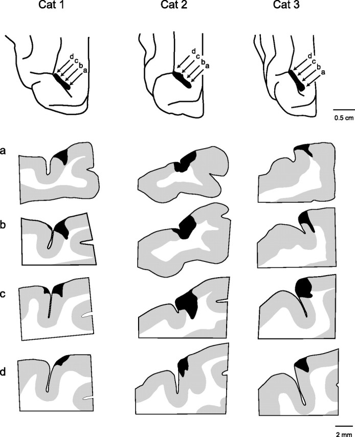
Figure 6.

Anatomy of lesions in area 5 of the right hemisphere of the three cats receiving simultaneous bilateral lesions (cats 1–3). The extent of the lesions in the posterior parietal cortex is shown at top of each column, and four cross-sections at different points in the lesion are shown in the columns. Rostral is to the left of each section. The locations of the cross-sections in rows a–d are indicated by the arrows in the top panels.
