
Figure 2.
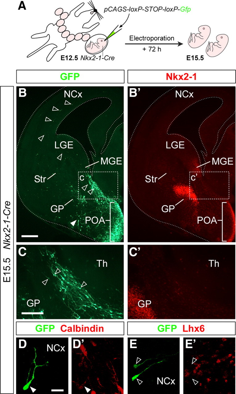
The embryonic POA gives rise to cells that migrate to the cortex. A, Schema of the experimental design followed in B–E′. B, B′, A representative case of the distribution of GFP (B)- and Nkx2-1 (B′)-expressing cells in a coronal section through the telencephalon of an E15.5 embryo in which the POA was electroporated at E12.5. The white arrowhead indicates the location of basal forebrain cells derived from the POA. C, C′, High-magnification images of the boxed areas shown in B and B′, respectively. The open arrowheads in B and C mark labeled cells with migratory morphology in the subpallium and in the cortex. D, D′, E, E′, Images of representative cells found in the cortex. These cells typically stain for Calbindin (D, D′) and do not express Lhx6 (E, E′). Scale bars: (in B) B, B′, 250 μm; (in C) C, C′, 100 μm; (in D) D, D′, E, E′, 25 μm.