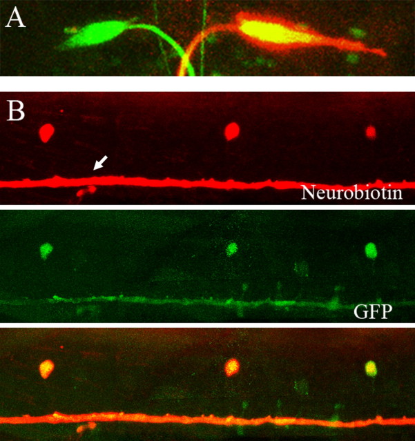
Figure 4.
Dye coupling between the Mauthner cell and CoLo neurons. Neurobiotin was loaded into one of the M-cells in a 3 dpf larva, and the sample was processed for Neurobiotin staining. A, In the hindbrain, one of the M-cells along with its axon shows Neurobiotin staining (red signal). B, In the spinal cord, CoLo neurons as well as the M-axon (arrow) show Neurobiotin staining, indicating that the CoLo cells are connected to the Mauthner axon via gap junctions.

