
Figure 7.
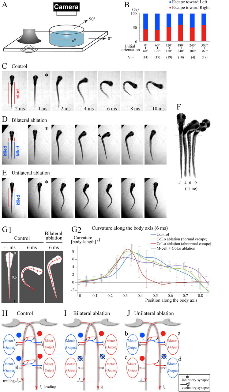
Behavioral analyses of the CoLo-ablated larvae. A, A schematic illustration of the experimental setup. Sound/vibration generated in the audio speaker propagates to the Petri dish. This elicits an escape in a larva. B, Relationships between the initial orientation of the larvae (see the schematics in A) and the direction of the escape. “N” stands for the number of the trials. The number of larvae used in this experiment was 16. C–E, Escape responses of a control (C) and CoLo-ablated (D, E) larvae. In each panel, a single asterisk marks the initiation of movement. Images were collected at 1000 frames/s. Every other frame is shown (2 ms between frames). C, An example of an escape behavior in a control larva (a Tol-056 heterozygote). See also supplemental Movie 3 (available at www.jneurosci.org as supplemental material). D, An example of an abnormal escape behavior in a CoLo-ablated larva (bilateral ablation of CoLos located caudal to the 7th segment). See also supplemental Movie 4 (available at www.jneurosci.org as supplemental material). Note that the caudal part of the body is almost straight, whereas contraction of the rostral part of the body seems normal. This resulted in a sharp kink (arrowhead). E, An example of an abnormal escape behavior in a CoLo-ablated larva (ablation of the right-side CoLos located caudal to the 7th segment). See also supplemental Movie 6 (available at www.jneurosci.org as supplemental material). Note that, near simultaneously, the rostral part of the body curved toward the right, whereas the caudal part of the body curved toward the left. This resulted in a characteristic S-shape of the body. F, Images of the bilaterally ablated larva at four different time points (the same trial shown in D). The onset of movement is defined as time 0. The pigmentation pattern was used to align larvae (the vertical line). Note that the straight part of the body is shortened. G1, Midline tracing of larvae. Images were inversed, and the midline was traced. Position along the body axis is indicated by bars at increments of 0.1. Left, A control larva before movement; middle, a control larva at 6 ms after the onset of movement; right, a bilaterally CoLo-ablated larva at the 6 ms time point. G2, Quantitative analyses of the curvature of the body at the 6 ms time point. Curvature of the midline at 0.001 increments of body length was calculated with BohBoh software. The blue trace represents the mean value of the escapes of control larvae. The green trace represents the mean value of the normal escapes of bilateral CoLo ablations. The red trace represents the mean value of the abnormal escapes of bilateral CoLo ablations. The purple trace represents the mean value of the escapes in those larvae in which unilateral ablation of the M-cell and bilateral ablation of CoLos were performed. The numbers of images examined are as follows: 14 for control; 18 for bilateral ablation with normal escapes; 9 for bilateral ablation with impaired escapes; 11 for unilateral M-cell ablation and bilateral CoLo ablation. For every 0.05 increment of position, the SDs are indicated. H–J, Schematic illustrations for explanation of the motor activities in control (H), bilaterally ablated (I), and unilaterally ablated (J) larva during coactivation of the two M-cells with slight time differences. In each panel, the leading spike runs on the right M-axon. Circular cells represent CoLo neurons. Red represents an active state, whereas blue represents an inactive state. H, The motor activity on the left side along the entire body is suppressed by the crossed inhibitions mediated by CoLos, resulting in a normal escape toward the right. I, In the rostral part of the body in which CoLos are present, the motor activity of the left side is suppressed. In the caudal part in which CoLos are absent, both sides become active, resulting in the straightening of the body. J, In the rostral part in which CoLos are present, the right side is active (a), whereas the motor activity of the left side is suppressed by CoLos (b). In the caudal part, in which CoLos are absent on the right side, the activity occurs on the left side (c). Activation of the right side depends on the balance between the excitatory effects originating from the right M-spike and the inhibitory effects originating from contralateral CoLos. If the inhibitions arrive early enough, the excitatory effects are suppressed (d).