Figure 1.
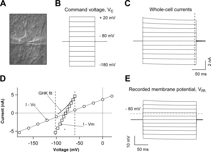
Characteristics of astrocyte passive conductance. A, DIC image of an astrocyte in the CA1 stratum radiatum during single-astrocyte dual-patch whole-cell recording, with two recording electrodes (shadows) sealed on the cell somata. While the conventional whole-cell recording was performed in one of the electrodes (the left electrode in A), the actual membrane potential (VM) change following the delivery of command voltages was recorded by the second electrode simultaneously (right electrode in A). B, Command voltage pulse protocol: 200 ms test voltages were stepped from −180 mV to +20 mV with an increment of 20 mV from a holding potential of −80 mV. C, E, The whole-cell currents recorded from the left electrode and actual membrane potentials VM recorded from the right electrode, respectively. D, Superimposing the current–command voltage relationship (I–VC) and current–membrane potential relationship (I–VM) from the same dual-patch recording revealed an ∼80% deficit of VM from the expected VC in voltage-clamp recording. The outward rectifying GHK currents generated by using the same chemical gradient of K+ in the recording solutions ([K+]i/[K+]o = 140/3.5 mm) is also shown in E, indicating a considerable deviation of the passive conductance from the expected GHK constant field rectification. As the GHK fit assumed only membrane permeability for the K+ ion, the GHK fit yielded a reversal potential of −93 mV, differing from the actual reversal potential of −87 mV measured from the dual-patch-recorded astrocyte.

