
Figure 9.
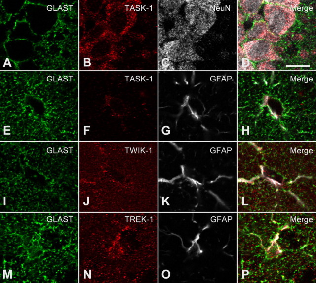
Immunocytochemistry of TASK-1, TWIK-1, and TREK-1 channel proteins in hippocampal sections. Coronal hippocampal sections were immunofluorescently labeled for one of the three K2P channel proteins, namely, TASK-1 (B, F), TWIK-1 (J), and TREK-1 (N), GLAST, (A, E, I, M), and NeuN (C) or GFAP (G, K, O). Shown are single confocal planes taken in the CA1 pyramidal cell layer (A–D), or the stratum radiatum (E–P). TASK-1 immunoreactivity was present in NeuN-immunoreactive pyramidal neurons (A–D) and to a lesser extent in GFAP- and GLAST-immunoreactive astrocytes (E–H). Note that the TASK-1 immunoreactivity overlaps with that for GFAP and is within the GLAST outline of the astrocyte soma (E–H). Compared with anti-TASK-1, anti-TWIK-1 and TREK-1 bound more in the neuropil, in a similar manner to anti-GLAST. These antibodies also labeled the GFAP(+) somata (see merged images L and P). The scale bar in D represents 10 μm and applies to all images in this figure.