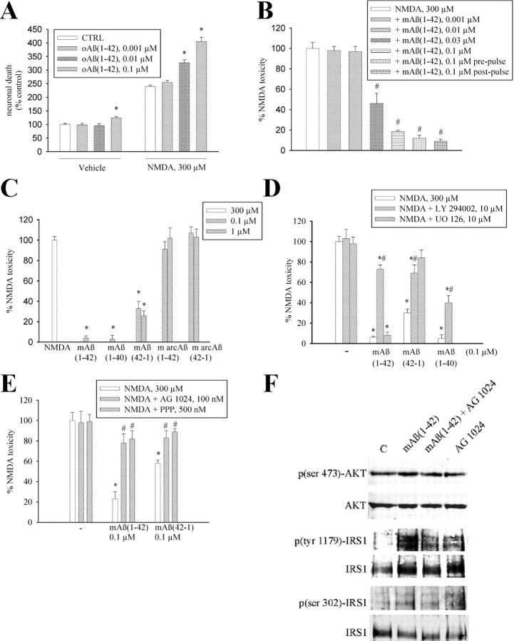
Figure 3.
Aβ1-42 monomers protect neurons against NMDA toxicity. A, NMDA-induced toxicity in mixed cortical cultures is potentiated by oligomeric Aβ1-42 [oAβ(1-42)] and (B) prevented by monomeric Aβ1-42 [mAβ(1-42)]. Toxicity was induced by a 10 min pulse with 300 μm NMDA and assessed by trypan blue staining (set to 100%) 24 h later. In the prepulse condition, Aβ1-42 monomers were applied 24 h before the NMDA pulse and cultures were extensively washed before the experiment. In the postpulse condition, Aβ1-42 monomers were applied soon after the excitotoxic pulse and kept for 24 h into the medium. Both in A and in B, values are means ± SEM of 9–18 determinations from three-six independent experiments. A, *Significantly different from the respective control (CTRL) at p < 0.05 (one-way ANOVA + Fisher's LSD test). B, *Significantly different from NMDA at p < 0.001 (one-way ANOVA + Fisher's LSD test). C, NMDA-induced toxicity in mixed cortical cultures is attenuated by the monomeric forms of Aβ1-42, Aβ1-40, and Aβ42-1, but not by Aβ1-42 or Aβ42-1 containing the Arctic mutation (m arcAβ). Values are means ± SEM of 12–18 determinations from three-six independent experiments. *Significantly different from NMDA at p < 0.001 (one-way ANOVA + Fisher's LSD test). D, The PI-3-K inhibitor LY 294002 (10 μm) prevents the neuroprotective activity of monomeric forms of Aβ1-42, Aβ1-40, and Aβ42-1. Values are means ± SEM of 8 determinations from two independent experiments. *,#Significantly different from NMDA (*) or from the respective Aβ conditions (#) at p < 0.05 (one-way ANOVA + Fisher's LSD test). [UO126] = 10 μm. E, The selective inhibitor of the insulin receptor superfamily, AG1024, and the preferential IGF-1 receptor inhibitor, picropodophyllin (PPP), prevent the neuroprotective activity of monomeric forms of Aβ1-42 and Aβ42-1. Values are means ± SEM of eight determinations from two independent experiments. *,#Significantly different from NMDA (*) or from the respective Aβ conditions (#) at p < 0.05 (one-way ANOVA + Fisher's LSD test). F, Representative immunoblots of p(ser 473)-AKT, p(tyr 1179)-IRS1, and p(ser 302)-IRS1 in pure neuronal cultures treated with 0.1 μm monomeric Aβ1-42 [mAβ(1-42)] for 5 min both in the absence and in the presence of 100 nm AG1024. Quantitation of p(ser 473)AKT/AKT ratios was as follows: control (C) = 0.5 ± 0.04; mAβ1-42 = 1.37 ± 0.1*; mAβ1-42 + AG1024 = 0.7 ± 0.08**; AG1024 = 0.55 ± 0.06 [means ± SEM of three independent experiments; *,**significantly different from control (*) or mAβ1-42 alone (**) at p < 0.05 by one-way ANOVA + Fisher's LSD test]. Quantitation of p(tyr 1179)-IRS1/IRS1 ratios was as follows: control (C) = 0.42 ± 0.12; mAβ1-42 = 1.31 ± 0.03*; mAβ1-42 + AG1024 = 0.63 ± 0.02**; AG1024 = 0.65 ± 0.08 [means ± SEM of three independent experiments; *,**significantly different from control (*) or mAβ1-42 alone (**) at p < 0.05 by one-way ANOVA + Fisher's LSD test]. Quantitation of p(ser 302)-IRS1/IRS1 ratios was as follows: control (C) = 0.39 ± 0.09; mAβ1-42 = 0.85 ± 0.06*; mAβ1-42 + AG1024 = 0.28 ± 0.11**; AG1024 = 0.46 ± 0.08 [means ± SEM of three independent experiments; *,**significantly different from control (*) or mAβ1-42 (**) alone at p < 0.05 by one-way ANOVA + Fisher's LSD test].

