Figure 1.
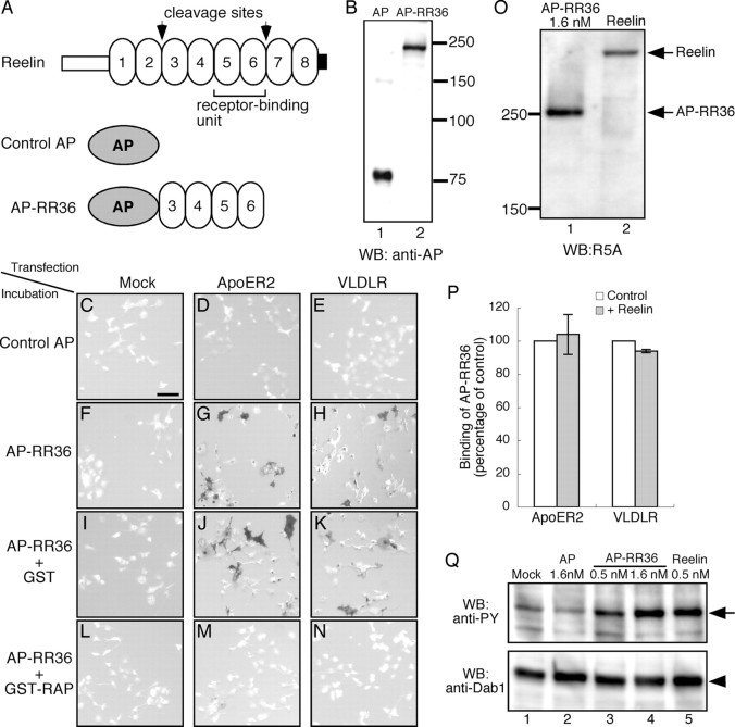
Construction of AP–RR36 and its ability to specifically detect FRRs. A, Schematic diagram of Reelin protein (top), control AP (middle), and AP–RR36 (bottom). Two sites that are cleaved in vivo by an unknown metalloprotease and minimum receptor-binding unit are shown in the top. B, Western blotting (WB) analysis. Culture supernatants containing either AP (lane 1) or AP–RR36 (lane 2) were separated by SDS-PAGE and proteins were transferred to a polyvinylidene difluoride membrane. The membrane was then incubated with anti-AP antibody, followed by incubation with HRP-conjugated secondary antibody and detection with a chemiluminescence kit. Positions of molecular weight markers are shown on the right in kilodaltons. C–N, AP–RR36 specifically binds to Reelin receptors. COS-7 cells were transfected with control vector (C, F, I, L), ApoER2 expression vector (D, G, J, M), or VLDLR expression vector (E, H, K, N). Two days later, the cells were incubated with control AP (C–E), AP–RR36 (F–H), AP–RR36 mixed with GST protein (I–K), or AP–RR36 mixed with GST–RAP (L–N). Cells expressing ApoER2 or VLDLR were stained with AP–RR36 (G, H, respectively). This staining was not affected by addition of GST protein (J, K) but was virtually abolished by coincubation with GST–RAP (M, N). O, Estimation of Reelin concentration. AP–RR36 (lane 1, 1.6 nm as calculated from AP-activity assay) (Flanagan et al., 2000) and Reelin (lane 2) were analyzed by Western blot using R5A antibody as described above. P, COS-7 cells expressing ApoER2 (left) or VLDLR (right) were incubated with culture medium from mock-transfected (white bars) or Reelin-transfected (gray bars) cells in the presence of sodium azide for 20 min. They were then incubated with AP–RR36, and the amount of its binding was quantitated as described in Materials and Methods (n = 3). Q, AP–RR36 induces phosphorylation of Dab1. Primary cortical neurons were incubated with the samples indicated above the lanes for 20 min at 37°C, and Dab1 phosphorylation was measured as described previously (Nakano et al., 2007). PY, Phosphotyrosine. Scale bar (in C): C–N, 100 μm.

