Figure 2.
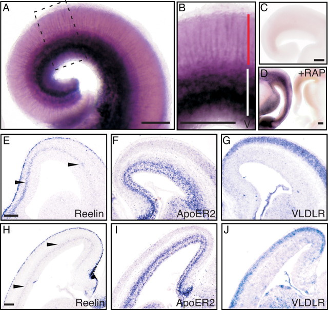
Localization of FRRs in the developing mouse cerebral cortex. A, B, E15.5 mouse brains were cut coronally and stained with AP–RR36. A magnified view of the area surrounded by a broken line in A is shown in B. Red and white bars in B indicate the region corresponding to the CP and IZ/SVZ, respectively. C, The brain slice from the same mouse as in A was stained by control AP. D, The staining by AP–RR36 is abolished in the presence of GST–RAP. Slices from an E15.5 mouse brain were stained with AP–RR36 in the presence of control GST (left) or GST–RAP (right). E–J, Localization of mRNA in the E15.5 cerebral cortex. Coronal (E–G) or sagittal (H–J) slices were hybridized with antisense probes for Reelin (E, H), ApoER2 (F, I), or VLDLR (G, J). Note the diffuse Reelin mRNA signal around the SVZ and IZ (arrowheads in E, H). Most of the ApoER2 mRNA is localized in the VZ/SVZ/IZ (F, I), whereas the VLDLR mRNA is more abundantly expressed in the CP (G, J). Scale bars: A–D, E (for E–G), and H (for H–J), 200 μm.

