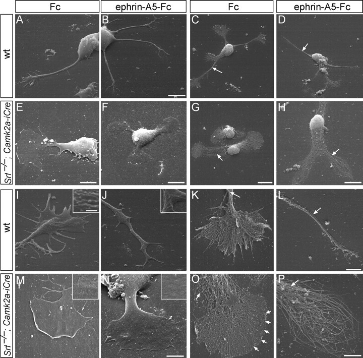
Figure 1.

Growth-cone inflation and absence of filopodia in SRF-deficient neurons. Neurons were fixed (A, B, E, F, I, J, M, N) or peeled off the plasma membrane (C, D, G, H, K, L, O, P) followed by platinum replica EM. A–D, Wild-type neurons protruded multiple long neurites terminated by growth cones with several filopodia (A, C), which after ephrin-A treatment collapsed (B, D). Note the tight filament bundling in the neurite shaft (C, D, arrows). E–H, Srf−/− neurons were stunted in growth, typically bipolar in shape and growth-cone area almost equaled cell-body area. Ephrin-A did not collapse growth cones (F, H) and produced ring-shaped filaments (H; see P). I–L, Wild-type growth cones contained filopodia separated by lamellipodia (E, K). Ephrin-A incubation reduced growth-cone complexity (J, L). Arrows in K and L point at tightly bundled filaments. The plasma membrane contained many ruffles and vesicle-like structure (J, K, insets). M–P, SRF-deficient growth cones (M, O) were rounded up and inflated with no distinguishable filopodia (O, arrowheads). Ephrin-A5 did not break down the cytoskeleton completely and resulted in ring-like filaments (N, P). Arrows in O and P point at loosened filament bundles. The plasma membrane appeared smooth (M, O, insets). Scale bars: A, B, 6 μm; C, D, G, 10 μm; E, 5 μm; F, H, 8 μm; I–K, M–P, 3 μm; L, 2 μm; insets, 500 nm.
