
Figure 2.
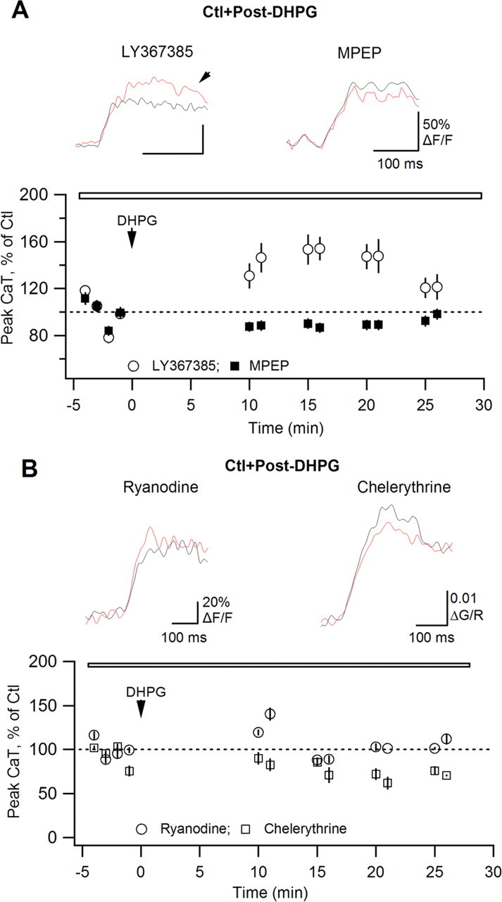
AP–CaT potentiation requires mGluR5, ryanodine-sensitive Ca2+ release and PKC activation. A, B, Summary plots of CaT peak amplitude versus time demonstrating the effects of DHPG on AP–CaTs in the presence of the mGluR1a antagonist LY367385 (A; 100 μm; n = 5), the mGluR5 antagonist MPEP (A; 5 μm; n = 5), the Ca2+ store inhibitor ryanodine (B; 30 μm; n = 5), and of PKC blocker chelerythrine (B; 10 μm; n = 4). Traces above are average expanded AP–CaTs (n = 3 traces) obtained in control (black) and after DHPG application (red).