
Figure 6.
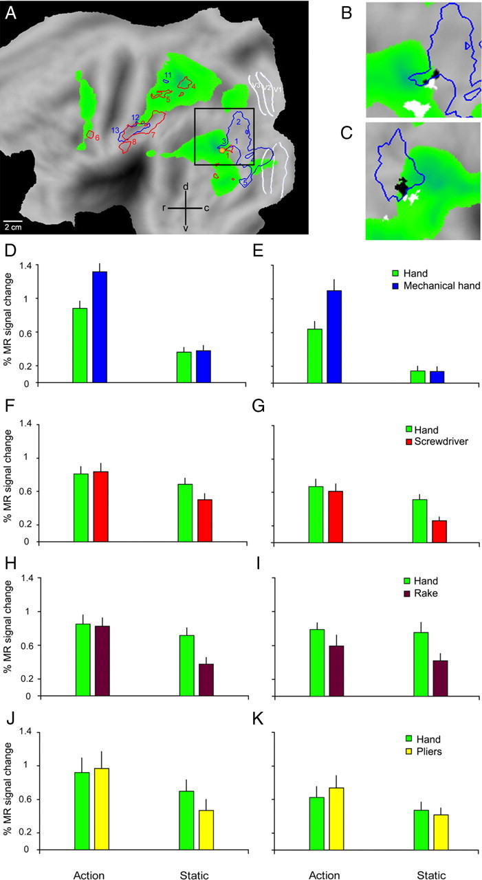
Human IOG interaction sites. A, Flatmap of the left hemisphere (human PALS atlas). Green areas show activations in the contrast observation of hand action minus static control (p < 0.001, experiment 1). Blue and red outlines show the regions where the interactions tool versus hand, relative to their controls, were significant in experiments 1 and 2 respectively (p < 0.001). White lines indicate areas V1-3; yellow dot, hMT/V5+; numbers indicate regions as listed in supplemental Tables S2 and S3, available at www.jneurosci.org as supplemental material; black square, part shown in B. B, C, Part of flatmaps of left and right hemispheres of the human PALS atlas showing the regions corresponding to the conjunction of the interactions for the 3 tools (white voxels), to the interaction for the mechanical hand (blue outlines), and the conjunction of the four interactions (black voxels). These latter voxels, located in IOG, reflect the overlap between two largely distinct regions, probably induced by the smoothing and the averaging across many subjects; green voxels same as in A. D–K, Activity profiles of left (D, F, H, J) and right (E, G, I, K) IOG regions yielded by the conjunction analysis of experiments 1–4 (black voxels in B, C). The larger percentage signal changes in these IOG regions compared with those in IPL (Figs. 3, 5) underscore their visual nature.