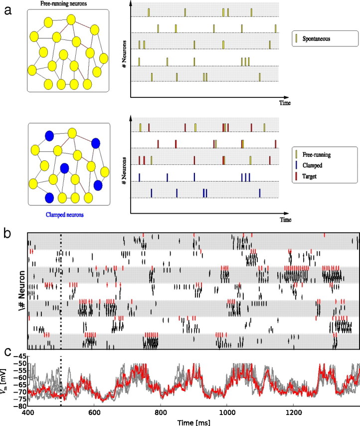
Figure 1.

a, Conceptual schema of the “frozen paradigm.” A spontaneous pattern is recorded (top panel), and then a subset of neurons (labeled “frozen”; blue neurons and blue spikes) is forced to replay part of the pattern. We then examined whether the remaining, free-running neurons (yellow spikes) reliably reproduce the other part of the spontaneous pattern, which we label the “target” pattern (red spikes) (see Results for details). b, Raster plot of responses of free-running neurons to the frozen stimulation. Each white or gray band represents the activity of one free-running neuron. Short vertical lines represent spikes. The red spikes are from the target pattern, and each row of black spikes represents a different trial with the same stimulation pattern but a different initial state of the network. c, Superimposed Vm traces of responses to the same frozen stimulation, for one free-running neuron. The red trace indicates the target activity.
