Figure 1.
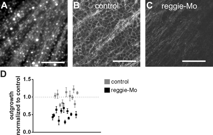
Reggie downregulation reduces axon outgrowth. A, Mo antisense oligonucleotides labeled with lissamine were applied immediately after ONS. A maximum-intensity projection of a deconvoluted Z-stack from a retina, 3 d after ONS, illustrates that RGCs and axons are labeled by retrograde Mo transport. B, C, Reggie-2 Ab immunostaining is present in control Mo-treated retinas which did not alter reggie expression (B). Reggie Mos reduced reggie-2 Ab staining of RGCs 7 d after ONS (C, images were taken with the same microscope settings). Scale bars, 50 μm. D, Regeneration was assessed by quantifying axons from mini-explants isolated 4 d after ONS. Retina pairs (21) (control- and reggie–Mo-treated eyes from the same fish) were analyzed. The mean number of axons per explant was normalized to the control retina, and relative outgrowth efficiency is shown.

