
Figure 3.
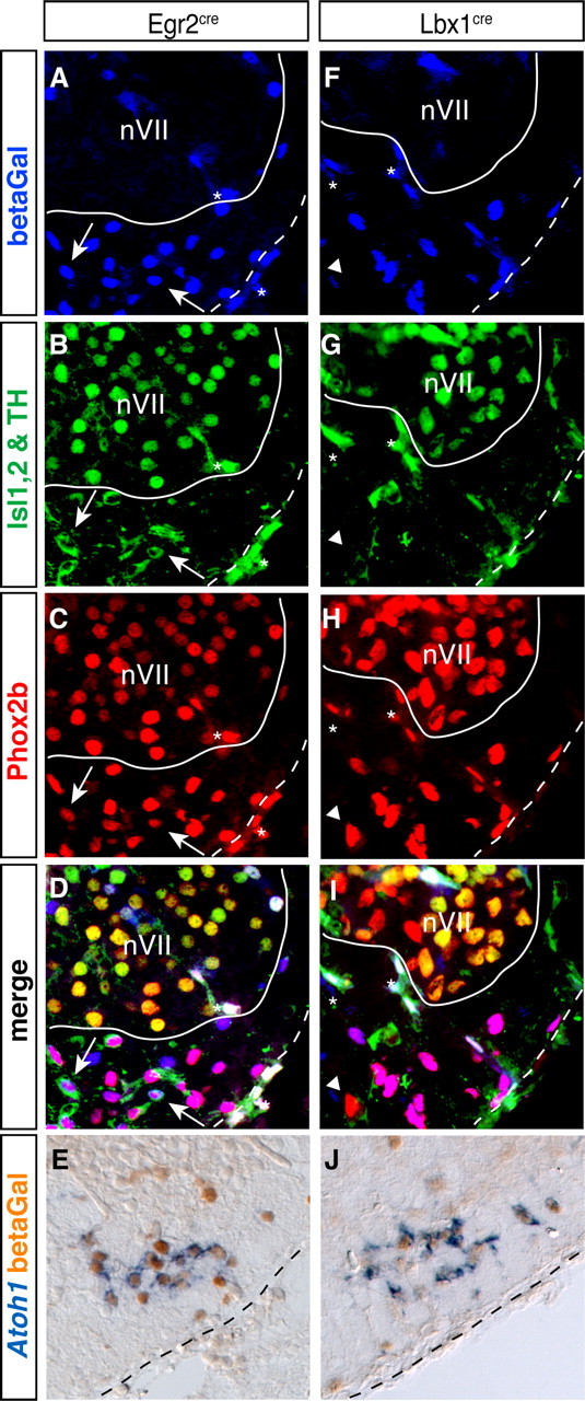
RTN neurons originate from Lbx1- and Egr2-expressing precursors. A–J, Transverse sections through E15.5 brains of Egr2cre/+; TauGFPnLacZ (A–E) and Lbx1cre/+; TauGFPnLacZ (F–J) embryos. A–D, F–I, Anti-β-galactosidase (blue, A and F), combined anti-Islet1,2 (nuclear labeling) and anti-TH (cytoplasmic labeling) (green, B and G), and anti-Phox2b (red, C and H) immunofluorescence. A white line delimits nVII, and a dotted line marks the ventral medullary surface. There are a few β-galactosidase+;Phox2b+;TH+ cells in the RTN from Egr2cre/+; TauGFPnLacZ embryos (white arrows) and one Phox2b+; TH−;Islet1/2− cell that is β-galactosidase-negative in the RTN from Lbx1cre/+; TauGFPnLacZ embryo (white arrowhead). Asterisks, nonspecific signal from blood vessels. E, J, ISH with an Atoh1 probe (blue) followed by anti-β-galactosidase immunohistochemistry (orange). A black dotted line marks the ventral medullary surface, lateral is to the right.