
Figure 4.
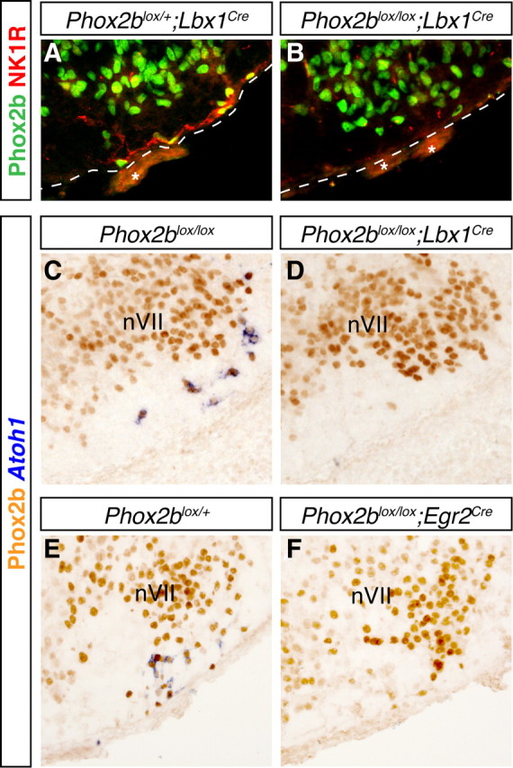
Loss of RTN neurons in Phox2b conditional null mutants. A, B, anti-Phox2b (green) and anti-NK1R (red) immunofluorescence on transverse sections through E15.5 brains of Phox2blox/+; Lbx1cre/+ (A) and Phox2blox/lox; Lbx1cre/+ (B) embryos. NK1R-positive cells and fibers at the ventral medullary surface beneath nVII are absent in the mutant embryo (B). A dotted line marks the ventral medullary surface. Asterisks, Nonspecific signal from blood vessels. C–F, ISH with an Atoh1 probe (blue) and anti-Phox2b immunohistochemistry (orange) on transverse sections through E15.5 brains of control (C, E), Phox2blox/lox;Lbx1cre/+ (D), and Phox2blox/lox; Egr2cre/+ (F) conditional mutants. Note the absence of Atoh1 staining in the conditional null mutants (D, F).