Figure 1.
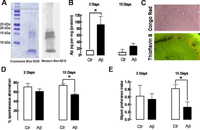
Intracerebroventricular administration of soluble β-amyloid peptides leads to an accumulation of soluble but not aggregated forms of Aβ in the hippocampus, causing delayed memory impairment without evident acute effects. The Coomassie R-250 staining and 6E10 antibody-based Western blot analysis of the two different batches of Aβ1-42 used showed that they were mainly constituted by monomers and oligomer containing up to four monomers (A). Rats were treated with Aβ1-42 (2 nmol, i.c.v.) or water (control), which accumulated in the hippocampus after 2 and 15 d (B), as measured by ELISA (n = 4 rats treated with water and n = 6 treated with Aβ1-42). Congo Red and Thioflavin S staining (C) failed to reveal the presence of Aβ aggregates in hippocampal sections collected 15 d after Aβ1-42 administration (images representative of 3 animals). D, Spontaneous alternation in the Y-maze test of control and Aβ1-42-treated rats after 2 or 15 d (n = 6 animals treated with water and n = 9 treated with Aβ1-42). E, Object recognition index in the object recognition test of control and Aβ1-42-treated rats after 2 or 15 d (n = 4 animals treated with water and n = 6–7 animals treated with Aβ1-42). Data in bar graphs are mean ± SEM; *p < 0.05.

