
Figure 2.
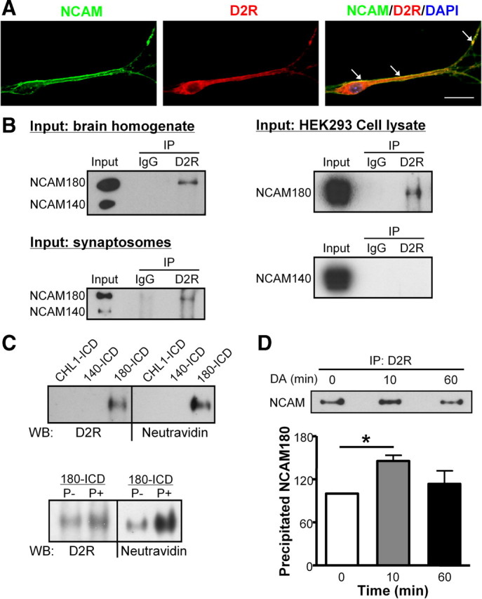
Interaction between NCAM and D2R in cells and tissue. A, Cultured hippocampal neurons were subjected to immunostaining with NCAM antibody and D2R antibody; nucleus was stained with DAPI. Colocalization of NCAM and D2R in cell soma and neurites is indicated by the white arrows. Scale bar, 20 μm. B, Brain homogenate, synaptosomal fraction, or HEK293 cell lysate transfected with D2R and NCAM were subjected to immunoprecipitation (IP) using mouse anti-D2R antibody or nonspecific mouse IgG. Precipitated proteins were analyzed by Western blot with NCAM antibody 5B8. Coimmunoprecipitation of NCAM180 and D2R was observed. C, NCAM180-ICD, NCAM140-ICD, or CHL1-ICD were coupled to the biotin containing cross-linker Sulfo-SBED and incubated as baits with brain homogenate under dephosphorylation (P−) or phosphorylation (P+) conditions. After UV cross-linking and denaturation under reducing conditions, the biotin moiety was transferred from the cross-linker to the molecules bound to the baits. Bound proteins were analyzed by Western blot (WB) using Neutravidin and anti-D2R antibody. D, Myc-D2R-expressing HEK293 cells were transfected with NCAM180 and stimulated with 10 μm DA for 0, 10, or 60 min. Cell lysates were prepared and subjected to immunoprecipitation (IP) using mouse anti-D2R antibody. Precipitated proteins were analyzed by Western blot with NCAM antibody 5B8. Coimmunoprecipitation of NCAM180 and D2R was observed. The amount of coimmunoprecipitated NCAM was quantified and the amount obtained after 0 min stimulation was set to 100%. Means + SEM are shown. *p < 0.05 by unpaired t test (n = 3).