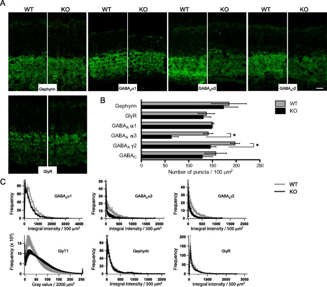
Figure 4.
GABAA receptor clustering is altered in the NL2-deficient retina. A–C, Representative single-plane confocal micrographs (A), numbers (B), and fluorescence intensity histograms (C) of puncta immunoreactive for gephyrin, glycine receptors, and GABAA α1, α3, or γ2 receptor subunits in the IPL of WT and NL2-deficient retinas (n > 6 mice). Whereas gephyrin and glycine receptor (GlyR) clusters distributed normally, GABAA γ2 and GABAA α3 receptors subunits immunoreactive puncta were far less abundant in the NL2 KO retina. GABAAα1 clusters appeared fainter, albeit present in normal amount. Of note, GlyT1 labeling was dimmer in the WT as in the NL2 KO, as represented by the high occurrence of low-intensity values (see also Fig. 3D). Scale bar, 10 μm. *p < 0.05.

