
Figure 5.
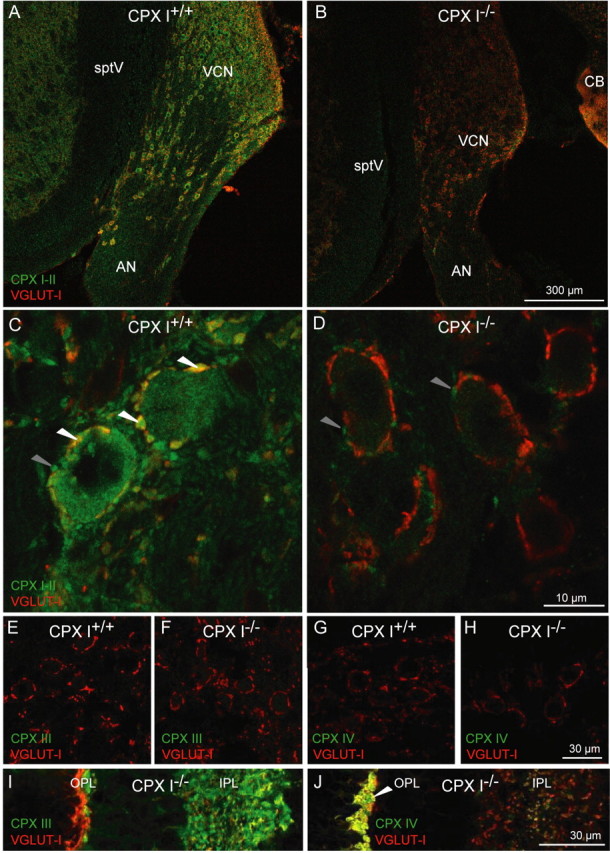
Distribution of CPX I and II protein in the cochlear nucleus. A, B, CPX I is expressed in the cochlear nucleus. Brainstem cryosections of 8-week-old mice were stained using antibodies against CPX I/II (green) and VGLUT1 (red). The VCN showed CPX I/II immunoreactivity in CPX I+/+ (A), but the staining was strongly reduced in CPX I−/− (B) sections. AN, Auditory nerve; CB, cerebellum; sptV, spinal tract of the trigeminal nerve; OPL, outer plexiform layer; IPL, inner plexiform layer. C, D, Higher-resolution confocal images of the auditory nerve entry zone into the cochlear nucleus of 8-week-old CPX I+/+ (C) and CPX I−/− (D) mice. In the wild type, CPX I/II immunofluorescent principal cells of the AVCN are engulfed by a ring-like array of CPX- and VGLUT1-positive spots (white arrowheads). In both CPX I+/+ and CPX I−/−, there were some CPX I/II-positive spots that were not costained with VGLUT1, presumably representing CPX II-expressing inhibitory synapses (gray arrowheads). The images in A,B and C,D were acquired at the same settings to illustrate the much-reduced CPX I/II immunofluorescence in the presynaptic and postsynaptic compartments of the CPX I−/− cochlear nucleus. E–J, Absence of CPX III and CPX IV in the cochlear nucleus. Brainstem cryosections of 8-week-old CPX I+/+ (E, G) and CPX I−/− (F, H) mice were stained using antibodies against CPX III (green; E, F) or CPX IV (green; G, H) and VGLUT1 (red). I, J, Cryosections of the retina of a CPX I−/− animal processed together with E–H and imaged with the same settings to demonstrate the absence of CPX III and IV immunofluorescence in the brainstem.