Figure 3.

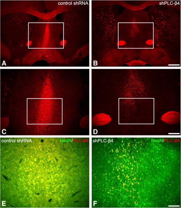
PLC-β4 expression in the medial septum of control shRNA mice and shPLC-β4 mice (A–D). Immunofluorescence staining for PLC-β4 in the medial septal area of shPLC-β4 mice is significantly downregulated (B, D). A normal PLC-β4 expression pattern is observed in the control shRNA mice (A, C). The rectangles in A and B indicate regions of C and D. Scale bars: A, B, 280 μm; C, D, 100 μm. Coexpression of NeuN and PLC-β4 in control shRNA mice and shPLC-β4 mice (E, F). Immunofluorescence staining for PLC-β4 in NeuN-positive neurons is markedly reduced in the medial septum of shPLC-β4 mice (F), whereas its expression in NeuN-positive neurons of control shRNA mice is generally unchanged (E). The rectangles in C and D indicate regions of E and F. Scale bar: E, F, 50 μm.
